CORNEA & CELL THERAPY
The cornea is the transparent dome shaped outermost part of the eye.
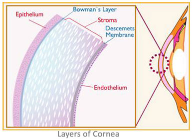
The cornea has five layers
- The epithelium
- Bowman’s Layer
- Stroma
- Descemet’s membrane
- Endothelium
 Epithelium
The epithelium is the outermost layer which serves as an effective barrier against the entry of foreign substances like dust, bacteria etc into the eye. It is composed of layers of non-keratinized stratified squamous epithelium in which the cells of the superficial layer are shed constantly and are replaced by the cells generated by multiplication in the basal layer. The epithelium is filled with thousands of tiny nerve endings that make the cornea extremely sensitive to pain when rubbed or scratched. The part of the epithelium that serves as the foundation on which the epithelial cells anchor and organize themselves is called the basement membrane.
The junction between the Cornea and the Sclera is called the Limbus and in this Limbus, the Corneal Epithelial Limbal stem cells are present. These stem cells when needed multiply and repair the damages of the corneal epithelial cell layer. When there is a damage to this Limbus itself (Eg stevens-johnson syndrome) or when the repairing capacity of the Limbal stem cells are overcome by the disease process (Eg. persistent corneal ulcer), damage to the corneal epithelium results in opacity affecting the vision.
There are two modalities of cell based therapies for the repair of the diseased corneal epithelium:
1. Limbal Tissue Based for unilateral corneal epithelial damage
2. Buccal Mucosa Based for bilateral cornal epithelial damanage